Quick and Easy Medical Terminologydisplacement of a Bone From a Joint is Called Quizlet
Overview

What is an elbow X-ray?
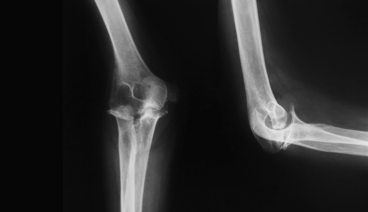
An elbow X-ray is a medical test that produces an image of the inside of your elbow. The image displays the inner structure (anatomy) of your elbow in black and white. An elbow X-ray shows your soft tissues and elbow bones. Your elbow bones include the upper bone of your elbow joint (humerus) and the lower bones of your elbow joint (radius and ulna). Your healthcare provider will use an elbow X-ray to diagnose and treat health and medical conditions in your elbow.
What are X-rays?
X-rays use a type of radiation called electromagnetic waves to create a picture of the inside of your body. Healthcare providers use X-rays more often than any other kind of medical imaging. X-rays send a beam of radiation through your body. Calcium in your bones absorbs more radiation, so your bones appear white. Soft tissues absorb less radiation, so they appear in various shades of gray. Air appears black.
When would I need an elbow X-ray?
Your healthcare provider will use an elbow X-ray to find the cause of any swelling, tenderness, pain or deformity in your elbow or elbows. They can use an elbow X-ray to diagnose possible health conditions involving your elbow. These conditions include:
- Broken bones (fractures) in your elbow.
- Dislocated elbow joints.
- Bursitis.
- Degenerative conditions such as arthritis in your elbows.
- Bone cysts.
- Bone infection (osteomyelitis).
- Bone cancer.
Your healthcare provider may use an elbow X-ray after a broken bone has been set. These X-rays can be used to make sure the bone was set properly and to ensure your bone or joint has healed correctly.
In addition, if you need elbow surgery, your healthcare provider will want X-rays before the procedure. They'll also want you to have routine follow-up X-rays afterward to track your condition.
Who performs an elbow X-ray?
A radiologic technologist (X-ray technician) will perform your elbow X-ray. Radiologic technologists are trained in patient care, radiation exposure, radiation protection, radiographic positioning and radiographic procedures.
Test Details
How does an elbow X-ray work?
X-rays send small beams of radiation through your body to create a picture. The picture is displayed on special photographic film or a digital platform.
Your body parts vary in thickness, so they absorb different amounts of radiation. Your bones absorb more radiation, so they look white on X-rays. Soft tissues such as your muscle, fat and organs are less dense, so they appear in different shades of gray. Air appears black.
How do I get ready for an elbow X-ray?
Elbow X-rays don't need a lot of preparation. You should wear comfortable clothing. You may have to remove your jewelry. Jewelry and other metal can show up on the X-rays. This can interfere with getting a usable image.
If you're pregnant, tell your radiologic technologist. Elbow X-rays use a very small amount of radiation and are considered safe during pregnancy. But your healthcare provider will decide if you need an X-ray. If it's urgent, your technologist will use precautions to reduce the risk of radiation exposure to your growing baby (fetus).
If you have any questions about the X-ray procedure, your technologist will be able to answer them for you.
What can I expect during an elbow X-ray?
A radiologic technologist will perform your elbow X-ray in your hospital's radiology department. You'll be led to a room with a large X-ray machine, where your technologist may give you a lead apron to protect you from radiation exposure. An X-ray is like getting a picture of your elbow taken — you can't feel it, and it creates an image. The procedure may take 15 minutes or more.
The technologist will have you place your arm on the X-ray table. They may put sponges or other positioning equipment around your elbow to keep it in place. You'll have to keep as still as possible during the test because any movement can affect the X-ray images. Your technologist will put an X-ray film holder or digital recording plate under the X-ray table. Then they will go into a small room or behind a wall to control the X-ray machine.
A normal elbow X-ray includes at least three images. Your technologist will return to reposition your elbow as needed. One image will be taken from the front (anteroposterior view), one image will be taken from the side (lateral view) and one image will be taken at an angle (oblique view). If you're in any pain, let your technologist know so they can help assist you through the test.
What can I expect after an elbow X-ray?
After your elbow X-ray, your radiologic technologist will want to make sure all of the images are clear. You'll be asked to wait while they check the images. If any of the images came out blurry, they'll have to retake them.
Then, a doctor called a radiologist will look over the images. Radiologists have special training in studying X-ray images and reading the results. Once the radiologist has read the results, they'll send them to your healthcare provider. Your healthcare provider will contact you to go over the results and discuss treatment.
Depending on the results, your healthcare provider may want you to return for a follow-up exam. They may need further X-rays of your elbow. They may also want you to return to track your condition over time.
What are the risks of an elbow X-ray?
X-rays are a quick and simple way for your healthcare provider to diagnose possible medical conditions in your elbow or elbows. Elbow X-rays give off only a small amount of radiation that goes directly through your body. X-rays don't cause side effects.
If you're pregnant, you have a slightly higher risk of problems with radiation exposure. Make sure to tell your radiologic technologist if you're pregnant or think you could be pregnant. Your technologist may give you a lead apron to protect your body from radiation exposure. Children also have a slightly higher risk. Your child's technologist may be able to use lower amounts of radiation.
Excessive amounts of exposure to radiation carries a small risk of cancer. If you're concerned about radiation exposure, talk to your technologist.
Results and Follow-Up
When will I find out the results of my elbow X-ray?
The results of your elbow X-ray may be available almost immediately if it was due to an emergency. Otherwise, your radiologist can usually get the results to your healthcare provider within one to two days. Your healthcare provider will then discuss the results with you.
Additional Details
Does tennis elbow show up on X-rays?
Tennis elbow is an overuse injury that occurs when your tendons become overloaded. This can lead to inflammation, degeneration and tearing. Tennis elbow can usually be diagnosed by a physical exam alone. X-rays don't clearly show the tendons in your elbow. However, your healthcare provider may order an elbow X-ray to rule out a fracture, dislocated joint or arthritis.
A note from Cleveland Clinic
X-rays are the most common type of medical imaging used today. If you have pain, swelling or tenderness in your elbow, your healthcare provider may order an X-ray to figure out what's going on. An elbow X-ray is a quick, easy and painless procedure. Your radiologic technologist will explain the process and answer your questions. While radiation exposure holds a very small risk, the amount you receive during an elbow X-ray is minor. Once you have the correct diagnosis, your healthcare provider will get you set with the proper treatment.
Source: https://my.clevelandclinic.org/health/diagnostics/23507-elbow-x-ray